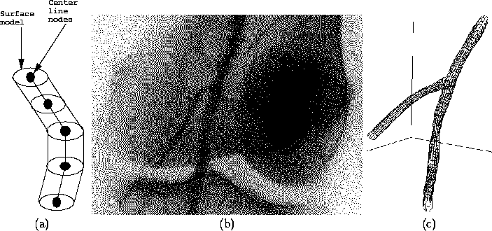
Figure 1: a) Model used to represent a blood vessel. b) An example angiogram image, showing arteries going through the knee. c) Reconstruction of the vessels shown in b.
Angiography is one of the methods used by radiologists to examine the structure and health of blood vessels. It consists of injecting an x-ray opaque contrast agent into the blood vessel, and taking one or more x-ray images of the vessel. While this method provides high resolution images, the data shows only two-dimensional structure. The goal of this project is to automatically reconstruct the three-dimensional structure of a blood vessel, using several angiogram images taken from different angles.
Among several types of information about blood vessels that are useful to physicians, the most important for diagnostic purposes is a measure of the cross-sectional area of the vessel at each location along its length. This information reveals narrow sections caused by blood clots, cholesterol build-up, or an injury. Treatment of these conditions involves inserting a catheter, with a small video camera and scraping tool mounted at its tip, into the vessel to remove the obstruction. A computer can aid in planning this surgery by simulating what the tip of the catheter would encounter in the vessel, and by computing how far the catheter must be inserted to begin work.
A semiautomatic system was developed to perform the three-dimensional reconstruction task. The representation used to model a vessel is shown in Figure 1a. First, to find the three-dimensional path of the center line of the vessel, we use a method called snakes [1]. Snakes are curves defined by a connected sequence of positions in space, each of which can move about in three dimensions. Each node experiences forces in the plane of the image which pull towards dark areas, to align curve with a blood vessel, and internal forces which keep the curve from developing sharp corners. By combining forces from several images taken at different angles, the snake is moved into alignment with the three-dimensional position of the vessel's center line.
The second step is to measure the diameter of the vessel. Just as a curve was used to model the center line of the vessel, a flexible cylindrical surface is used to represent the vessel's actual shape [3]. In each image, the program examines the intensity profile across the vessel, and matches this with an expected intensity profile [2]. From this, it is possible to estimate the location of the boundary of the vessel, and these estimates are used to apply forces to the flexible surface model.
The above system has been tested with angiogram images provided by Shadyside Hospital. Figure 1b shows one of a sequence of angiogram images, and Figure 1c presents the three-dimensional reconstruction of the main vessel and a branch coming off of it. Note that a narrow area in the main vessel, below the branch, has been successfully reconstructed.

Figure 1: a) Model used to represent a blood
vessel. b) An example angiogram image, showing arteries going through
the knee. c) Reconstruction of the vessels shown in b.
The system developed has so far been applied to two sets of angiogram images, and has been able to reconstruct the larger vessels and gross structure in each set. These models can be displayed by the computer from any viewpoint. However, more work is needed in a few areas.
The current program requires a significant amount of manual input to set the initial position estimates of the blood vessel center lines. These estimates need not be particularly accurate, as the snakes algorithm will refine the estimates. However, when attempting to reconstruct a large number of small vessels, such manual input can become quite tedious and error-prone. Current work involves further automating the process of finding vessel center lines, possibly through the use of algorithms originally developed for stereo vision.
Finally, for these reconstructions to be useful, they must be accurate. While it may not be possible to compare the model with direct physical measurements, it is possible to use other imaging modalities such as CT (Computed Tomography) and ultrasound to cross-check the results. We are working with Shadyside Hospital to perform such comparisons.