
Paper:
Z. Yin, K. Li, T. Kanade and M. Chen, "Understanding the Optics to Aid Microscopy Image Segmentation," the 13th International Conference on Medical Image Computing and Computer Assisted Intervention (MICCAI), 2010. [PDF]Abstract:
Image segmentation is essential for many automated microscopy image analysis systems. Rather than treating phase contrast microscopy images as general natural images and rushing into the image processing warehouse for solutions, we propose to study microscope's optical properties to model its image formation process first. It turns out that the phase contrast imaging system can be relatively well explained by a linear imaging model. Using this model, we formulate a quadratic optimization function with sparseness and smoothness regularizations to restore the "authentic" phase contrast image that directly corresponds to specimen's optical path length without phase contrast artifacts such as halo and shade-off. With artifacts removed, high quality segmentation can be achieved by simply thresholding the restored image. The imaging model and restoration method are quantitatively evaluated on two sequences with thousands of cells captured over several days.
Results:
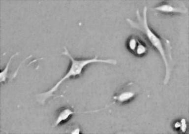
Input:


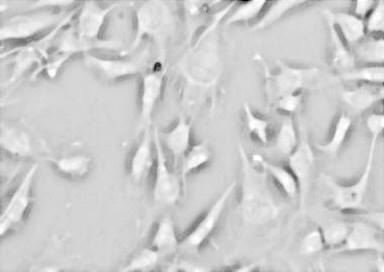
Restoration (videos):

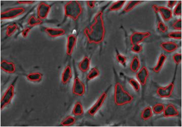
Segmentation:


More restoration processes (videos):

