Back to "Biological Language Modeling Seminar Topics"

Reference: www.mshri.on.ca/pawson/research1.html

Back to "Protein protein interactions"


Chromo Domains


Structure

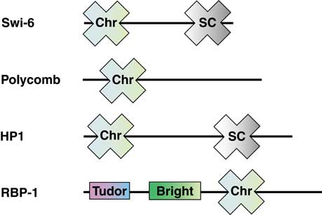
The Chromo domain appears as an N-terminal three-stranded anti-parallel b-sheet which folds against an N-terminal a-helix. A conserved series of hydrophobic residues that winds across the face of the b-sheet is referred to as a ‘sash’. Interactions with partner proteins are thought to be mediated by the residues within the hydrophobic sash. The figure shows the structure of the chromo domain from mouse modifier protein 1. Reference: Ball et al. 1997 EMBO J. 16:2473-2481.


Domain binding and function

The chromatin organization modifier (Chromo) domain is defined as a 30-70 amino acid residue protein module found in a number of proteins involved in the assembly of protein complexes on chromatin. This domain was first described in Drosophila modifiers of variegation, which are proteins that modify the structure of chromatin to the condensed morphology of heterochromatin, a cytologically visible condition where gene expression is repressed. Examples of Chromo domain containing proteins include the HP1 molecule involved in repression of gene expression in heterochromatin, the Polycomb (Pc) transcriptional repressors of homeotic genes in which the Chromo domain is essential for chromatin targeting, and human retinoblastoma binding protein (RBP1). Some Chromo domain (CD) proteins contain an N-terminal Chromo domain and a C-terminal Shadow Chromo domain (CSD).


Binding examples

Chromodomain proteins

Binding partner 

HP1 Histone H3 methylated lysine-9